
3D airway reconstruction for voice restoration
- MIRAI 3D
- 15 abr 2021
- 1 Min. de lectura

Dr. Alejandro Bertolotti
Hospital Universitario Fundación Favaloro - Buenos Aires, Argentina
Benefits
✔ Increased understanding of the lesion
✔ Design and optimisation of the surgical plan.
✔ Improved interaction between the surgical team
✔ Improved safety at the time of surgery
✔ Guidance in the surgical approach during the intervention
✔ Improved post-operative care and quality of life of the patient


Clinical case
A 30-year-old man was involved in a traffic accident that caused a traumatic brain injury and the destruction of his trachea and larynx.
The patient had to undergo emergency surgery, lost the sight in one eye and, due to the total occlusion of the larynx, required a tracheostomy with connection to mechanical ventilation. After he was stabilised, a second operation was scheduled to perform an airway plasty, where the 3D printed biomodel with his specific anatomy was used.


3D anatomical model
◾ FDM technology
◾ Material: PLA
◾ Resolution: 0.2 mm
◾ Finish: One colors
Surgical plan and results in the operating room


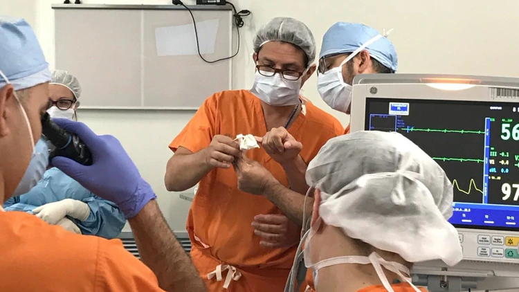


The doctor in charge, having already used 3D models in the past, requested that a biomodel be made to replicate the pathological situation presented by the patient and thus optimise the surgical strategy. Thus, together with the surgical team, they defined the laryngotracheal plastic reconstruction.
The three-dimensional model easily showed both healthy and affected areas of the airway and the anatomical relationship between them. During the operation, the doctors relied on the biomodel several times to perform the reconstruction and proceed with complete precision.
The use of the same for planning and guiding the surgery allowed for a successful procedure, and for the patient to regain speech after three months of tracheotomy.