
3D models for pancreatic hydatid cyst resection
- MIRAI 3D
- 27 abr 2021
- 2 Min. de lectura

Actualizado: 4 may 2021
Dr. Horacio Bignon, Dr. Adolfo Pereyra Rozas & Dr. Marcelo Muñoz
Sanatorio Nuestra Señora del Rosario - Jujuy, Argentina
Benefits
✔ Dimensioning the structures in 3D
✔ Evaluating the relationship between structures
✔ Designing the surgical plan
✔ Reduction of total surgery time
✔ Real-time guidance of surgical approach


Clinical case
An 11-year-old female paediatric patient with no history of illness.
She presented to the clinic for abdominal pain and they decided to perform an ultrasound scan. In the ultrasound, in addition to finding the reason for the complaint, they discovered a lesion in the left hypochondrium.
As a result of these findings, they decided to perform an abdominal CT scan to evaluate the lesion. A rounded formation was identified adjacent to the pancreatic tail and the splenic hilum, which was related on its posterior side to the left kidney and the left renal vein, on its upper side to the gastric body, on its lower side and on its anterior side to the distal end of the transverse colon. Its imaging appearance suggests in the first instance an intestinal duplication cyst, i.e. a congenital digestive anomaly.


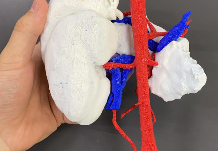
3D anatomical model
◾ FDM Technology
◾ Material: PLA
◾ Resolution: 0.2 mm
◾ Finish: Multiple colours
Surgical planning and results in the operating theatre
Dr. Bignon had a virtual 3D anatomical model and a physical one to plan the surgery. They noted that it was an abdominal cystic tumour and not a congenital anomaly, information that was corroborated after surgery with pathological anatomy.
Because the tumour was in close relationship with the aforementioned organs and their respective blood vessels, the biomodels allowed him to accurately visualise this relationship and assess the extent of the lesion.
The 3D models clearly showed that the tumour did not reach the splenic artery and vein so it was decided that the surgical plan to follow was a caudal pancreatectomy with preservation of the spleen. The approach chosen was laparoscopic. During surgery, due to the size of the tumour, it was decided to make a suprapubic incision to remove the tumour mass more safely.


Dr Bignon said that reconstruction and 3D printing "gives you a three-dimensional view of the surgery, gives you a roadmap and allows you to better understand where to approach the tumour" but also stressed that 3D models must be learned to use them, as from his own experience in this first case, he noticed that the model shows many details that he had not noticed until after seeing them in the operating theatre on the same patient. In other words, knowing how to use the biomodels and getting used to them allows one to have a large amount of information that results in making better decisions in advance and not in the operating theatre.
The 3D models also provided a reduction in surgical time, allowing the surgery to be performed in approximately 120 min.
Finally, the pathological anatomy categorised the abnormal mass as hydatidosis or hydatid cyst, which is a parasitic disease spread by contact with animal faeces contaminated with tapeworm eggs through water, contaminated food or animal skin.


Do you want to know more cases of BPH surgery? "Pancreatic tumour biomodel for paediatric surgery". At the Garrahan Foundation, Dr. Horacio Questa used a three-dimensional model to plan the resection of a Frantz's tumour in a 9-year-old patient.