
3D planning for adult congenital heart disease
- MIRAI 3D
- 26 abr 2021
- 2 Min. de lectura

Actualizado: 5 may 2021
Dr. Mariano Vrancic
Instituto Cardiovascular de Buenos Aires (ICBA) - Buenos Aires, Argentina
Benefits
✔ Detailed knowledge of the anatomy
✔ Taking measurements on the model
✔ Design of the operative plan
✔ Guidance in the surgical approach in the operating theatre
✔ Increased safety


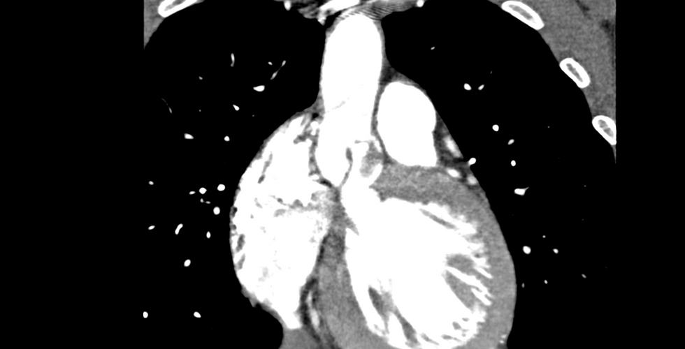


Clinical case
28-year-old female patient with a history of surgery for subaortic membrane at 4 years of age, reoperation at 10 years of age due to recurrence of the membrane and bicuspid aortic stenosis.
Given the recurrence of subaortic stenosis, the patient required a new operation to improve her heart's outflow.
3D anatomical model

Two physical biomodels were made:
1. Endocardial surfaces: printed with FDM technology in PLA material, red and solid, with a resolution of 0.2 mm.
2. Aortic valve: Printed with SLA technology in translucent and flexible resin, with a resolution of 0.02 mm.
What happened in the operating room after the 3D simulation?

Dr. Vrancic had multiple virtual 3D anatomical models, showing the complete endocardial surfaces, and with various slices (transverse or longitudinal) in order to perform a thorough assessment of the patient's anatomy.
In addition, as mentioned above, he used two physical biomodels. The first was rigid and corresponded to the endocardial surfaces of the upper half of the heart. The second was a flexible, translucent biomodel of the aortic valve.
The treatment was aortic valve replacement surgery with enlargement of the annulus and outflow tract of the left ventricle (Rastan-Konno surgery).
As this was a valve repair for a complex and rare pathology for adult cardiovascular surgeons, the 3D model allowed them to see the anatomy in detail and to programme the exact technique. They were able to measure the different diameters on the biomodel before surgery and after simulating the surgery.
Watch this video to see how 3D anatomical models are used for accurate and appropriate patient planning:
"It was very useful to understand the anatomy and to plan the type of surgery and eventually the necessary material", says Dr. Mariano Vrancic of his experience using biomodels.
The surgery was performed successfully and the patient is in good health and is close to being discharged from the hospital.
Discover more applications in cardiovascular surgery: "Simulation in vascular surgery: 3D training for catheterisation": Clinical case of Dr. Mariano Ferreira, at Clínica La Sagrada Familia, where they use 3D models to practice their interventions and train doctors in training.