
Thorax 3D - Narrowing resection margins and achieving more cost-effective surgery
- MIRAI 3D
- 3 may 2021
- 2 Min. de lectura

Actualizado: 4 may 2021
Dr. Matías Nicolás
Clínica Privada de Comunidad - Mar del Plata, Argentina
Benefits
✔ Accurately interpret the lesion
✔ Designing and optimising the surgical strategy
✔ Visualise and practise the plan to be followed on the model
✔ Guiding the approach during surgery
✔ Improve patient-physician interaction
✔ Enrich communication between involved clinicians
✔ Decrease the patient's post-operative time


Clinical case
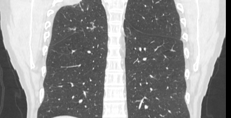
A 45-year-old man was diagnosed with a right upper lobe lung tumour. The tumour invaded the chest wall and the indicated treatment was resective surgery after chemotherapy and neoadjuvant radiotherapy.
For the planning of the surgery, a customised anatomical model of the patient was used to replicate the full-scale anatomy.


3D anatomical model
◾ FDM Technology
◾ Material: PLA
◾ Resolution: 0.2 mm
◾ Finish: Multiple colours
Surgical planning and outcomes in the operating room
Dr. Nicolás had a virtual and a physical 3D model that allowed him to clarify all the doubts about the anatomy and the extent of the anomalous mass that the two-dimensional images could not resolve. This gave him the possibility to optimise the resection margins.
Are you a specialist in thoracic surgery?
Don't miss lesson 4 of the free course Innovation in Surgery 3 on thoracic surgery this Thursday 6th May. Register here: https://www.en.modelosmedicos.com/webinar You will learn how you can improve your training using 3D planning and simulation.
The mini-invasive surgical plan chosen was a right upper lobectomy by uniportal video-assisted thoracoscopic surgery (UVATS) with rib resection. The physical biomodel was useful to visualise and project how the selected procedure would be performed for the patient.

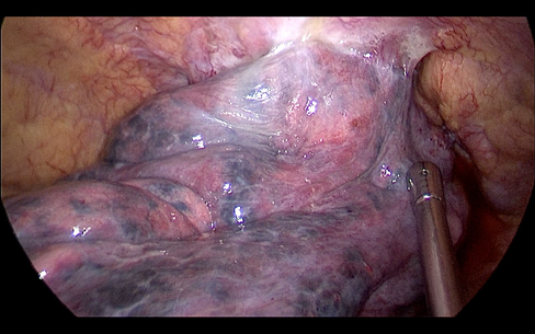


The 3D anatomical model was also used in the operating room, which was consulted several times to guide the approach to the tumour and was fundamental to recognise the repair points on the rib that delimited the resection, Dr Nicolás said.
In addition, the doctor used the 3D models to explain the surgery to the patient, showing him greater commitment and expertise in the case and thus transmitting security and peace of mind.
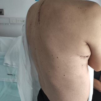
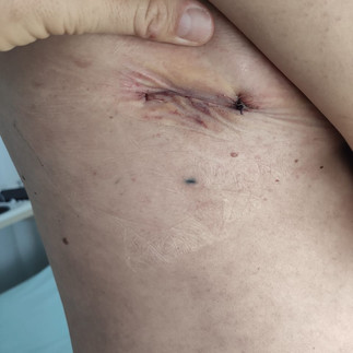
The surgery was successfully performed in 280 minutes and the patient showed a rapid and very favourable evolution.


Video of the surgery
You may be interested in: "Radical thymectomy with virtual 3D surgical planning". At Hospital Vozandes (Quito, Ecuador) Dr Guamán used a virtual 3D model to plan a radical thymectomy in a 67-year-old female patient.