
What are the benefits of 3D technologies for resecting tumours in the pancreas?
- MIRAI 3D
- 16 abr 2021
- 2 Min. de lectura

Dr. Federico Walter García
Clínica Pueyrredón - Mar del Plata, Argentina
Benefits
✔ Surgical plan design
✔ Reduction of surgical times
✔ Reduction of bleeding
✔ Guidance in the operating theatre approach
✔ Improved interaction between involved clinicians
Clinical case


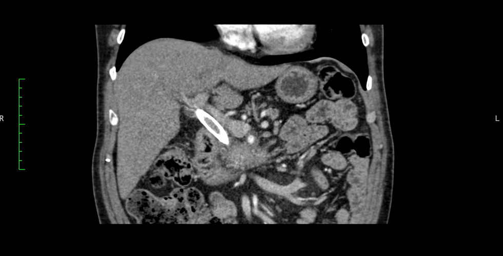


71-year-old woman diagnosed with resectable borderline pancreatic cancer (tumour of low malignant potential).
Taking into consideration the possibility of aggravation of the diagnosis, the attending physicians decided to perform a resection of the tumour located in the head of the pancreas.
Correct assessment of the tumour was of utmost importance because of its proximity to the duodenum, the superior mesenteric vein, the hepatic portal vein and the superior mesenteric artery.
Surgical plan and results in operating room


The surgical plan consisted of a cephalic duodeno-pancreatectomy, which was started with a laparoscopic approach and then converted to open surgery for safety when detaching the tumour from the portal vein and superior mesenteric vein.
A 3D virtual model with the patient's specific anatomy was used to design the resection strategy. This allowed the surgical team to optimise and reduce surgical times: ischaemia time and time in the operating theatre, resulting in a total procedure of 5 hours. Moreover, by knowing where they would find the vessels to be ligated, they were able to recognise in advance the critical moments of the surgery and reduce bleeding.

The surgery was successfully performed and the patient was discharged on the fifth day after the operation.
"The experience was very good, it allowed us to have a more realistic planning at the time of surgery", Dr. Federico Walter García concludes, after using 3D surgical planning for the first time.
Do you want to know more cases of HPB surgery? "Pancreatic tumour biomodel for paediatric surgery". At the Garrahan Foundation, Dr. Horacio Questa used a three-dimensional model to plan the resection of a Frantz's tumour in a 9-year-old patient.